
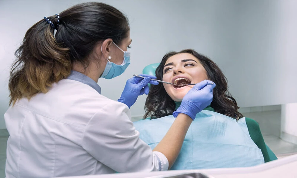

You sit back in the chair, the images go up on the screen, and your dentist in Lansing MI starts comparing today’s scans to ones from your last visit. That moment can make people tense. You may be wondering if they see something serious, if a small issue got worse, or if you somehow missed a warning sign. That reaction is common. Dental images can feel technical and distant, but to a general dentist, they tell a very practical story about what has changed, what has stayed stable, and what needs attention before it turns into pain or cost.
When a dentist compares current and previous dental images, they are not just looking for cavities. They are tracking patterns. They can spot bone loss, watch fillings and crowns for wear, check whether a suspicious area is active or unchanged, and see whether treatment is working. That side by side view often matters more than a single image on its own, because change over time is what reveals risk.
Comparing dental X-rays shows change that a single image cannot
A single dental image gives a snapshot. A series of images gives a timeline. That difference matters. A tiny shadow between two teeth may not mean much by itself, but if it was not there last year and is larger now, your dentist has stronger evidence that decay is progressing. The same is true for bone around teeth. Mild bone loss can look easy to overlook until you compare it to an earlier image and see that the support around the tooth is slowly dropping.
This is one reason dentists do not rely only on what they can see during an exam. Some problems grow under the surface. A tooth can look fine from above while decay moves between teeth or under an old filling. An infection can form near a root before swelling or sharp pain starts. Reviewing older images helps separate a stable finding from one that is becoming a problem.
You might hear your dentist mention “watching” an area. That can feel unsettling, as if nothing is being done. In many cases, watching is the right call. If a spot has not changed across several visits, immediate treatment may not be necessary. If that same spot expands, darkens, or starts affecting nearby structures, the plan changes because the evidence changed.
Past and present dental images help your dentist catch small problems early
Early detection often saves tooth structure, time, and money. A cavity found when it is small may need a simple filling. The same cavity, left to grow, can move into the nerve and lead to a root canal or extraction. Comparing images helps your dentist judge speed, not just presence. Is that crack line stable, or is it spreading? Is the bone around an implant holding firm, or is there slow loss that needs attention now?
This side by side review also protects you from overtreatment. A shape that looks odd on one image may simply be normal anatomy if it has looked the same for years. A dentist who sees no change may decide the safest choice is to monitor, not drill. That is one of the less obvious benefits of comparing dental X-rays over time. It can support treatment, and it can support restraint.
According to the FDA guidance on dental radiographic examinations, dental X-rays should be selected based on your needs rather than taken on a fixed schedule for every patient. That approach makes sense when you think about image comparison. The goal is not more images for the sake of it. The goal is useful images taken when they can answer a real clinical question.
What a general dentist looks for in old and new dental images
A general dentist usually compares images for a few core reasons. Decay is one of them, especially between teeth and under existing dental work. Bone levels are another, since they show whether gum disease is stable or getting worse. Existing restorations matter too. Fillings can leak, crowns can develop open margins, and root canals can show signs of healing or failure over time.
Growth and development can also be part of the picture. In younger patients, dentists may compare images to see how adult teeth are erupting and whether wisdom teeth are moving into a harmful position. In adults, they may track wear from grinding, changes in the jawbone, or signs of infection at the root tips.
If you want a plain language overview of how X-rays work and what they show, RadiologyInfo’s explanation of X-rays is helpful. For many patients, the stress drops once they understand that these images are not mysterious. They are simply a tool that lets your dentist see what eyes alone cannot.
Benefits and limits of comparing current and previous dental images
| What your dentist compares | What they may learn | Why it matters to you |
|---|---|---|
| Small dark areas in enamel or dentin | Whether decay is stable or growing | Early treatment may prevent a larger filling or root canal |
| Bone height around teeth | Whether gum disease is active | Intervening sooner may help preserve teeth |
| Margins around fillings and crowns | Whether old dental work is leaking or failing | Repairing problems early can avoid fractures or infection |
| Areas near root tips | Whether infection is healing, unchanged, or worsening | Treatment decisions become clearer and more precise |
| Suspicious but unchanged findings | Whether monitoring is safer than treatment | You may avoid unnecessary procedures |
People also worry about radiation, and that concern is reasonable. The good news is that dental X-ray exposure is low, and modern systems often reduce it further. The CDC’s facts about X-rays explain that medical imaging uses small amounts of radiation, with safety based on using the lowest amount needed to get useful information. That fits the same pattern your dentist follows when deciding if updated images are worth taking.
Three steps to take before and after dental imaging
Ask what changed since your last images. You do not need to interpret the picture yourself. Ask your dentist to point to the exact area and explain whether it is new, larger, smaller, or unchanged. That one question often clears up fear fast.
Keep your dental visits and records consistent. The value of current and previous dental images grows when they can be compared over time. If you change offices, request that your records be transferred so your new provider can see the pattern, not just the latest snapshot.
Follow through on monitoring plans. If your dentist recommends watching an area in six or twelve months, that does not mean the issue is minor enough to forget. Monitoring only works when you return on schedule. Missed follow up can turn a manageable issue into a painful one.
Dental image comparison gives you a clearer path forward
When your dentist studies old and new images together, they are building a timeline of your oral health. That timeline helps them find trouble early, avoid unnecessary work, and make treatment decisions based on evidence instead of guesswork. If you have ever felt uneasy when the images go up on the screen, you are not overreacting. You are seeing the part of dentistry where small changes matter, and where careful comparison often protects you from bigger problems later.
If your dentist recommends updated imaging or wants to review older scans with you, that is your chance to get clear answers and a plan you can trust.
What Your Dentist Learns From Comparing Current And Previous Dental Images